 W
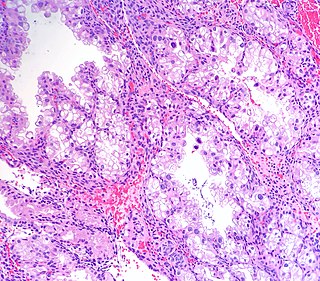
WArias-Stella reaction, also Arias-Stella phenomenon, is a benign change in the endometrium associated with the presence of chorionic tissue.
 W
WThe Bartholin's glands are two pea sized compound alveolar glands located slightly posterior and to the left and right of the opening of the vagina. They secrete mucus to lubricate the vagina and are homologous to bulbourethral glands in males. However, while Bartholin's glands are located in the superficial perineal pouch in females, bulbourethral glands are located in the deep perineal pouch in males. Their duct length is 1.5 to 2.0 cm and they open into navicular fossa. The ducts are paired and they open on the surface of the vulva.
 W
WThe broad ligament of the uterus is the wide fold of peritoneum that connects the sides of the uterus to the walls and floor of the pelvis.
 W
WThe cervical canal is the spindle-shaped, flattened canal of the cervix, the neck of the uterus.
 W
WThe cardinal ligament is a major ligament of the uterus. It is located at the base of the broad ligament of the uterus. There are a pair of cardinal ligaments in the female human body.
 W
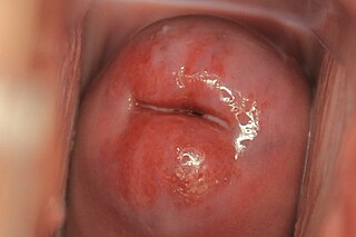
WCervical ectropion is a condition in which the cells from the 'inside' of the cervical canal, known as glandular cells, are present on the 'outside' of the vaginal portion of the cervix. The cells on the 'outside' of the cervix are typically squamous epithelial cells. Where the two cells meet is called the transformation zone, also known as the stratified squamous epithelium. Cervical ectropion can be grossly indistinguishable from early cervical cancer and must be evaluated by a physician to determine risks and prognosis. It may be found incidentally when a vaginal examination is done. The area may look red because the glandular cells are red. While many women are born with cervical ectropion, it can be caused by a number of reasons, such as:Hormonal changes, meaning it can be common in young women Using oral contraceptives Pregnancy.
 W
WThe clitoris is a female sex organ present in mammals, ostriches and a limited number of other animals. In humans, the visible portion – the glans – is at the front junction of the labia minora, above the opening of the urethra. Unlike the penis, the male homologue (equivalent) to the clitoris, it usually does not contain the distal portion of the urethra and is therefore not used for urination. The clitoris also usually lacks a reproductive function. While few animals urinate through the clitoris or use it reproductively, the spotted hyena, which has an especially large clitoris, urinates, mates, and gives birth via the organ. Some other mammals, such as lemurs and spider monkeys, also have a large clitoris.
 W
WThe corona radiata is the innermost layer of the cells of the cumulus oophorus and is directly adjacent to the zona pellucida, the inner protective glycoprotein layer of the ovum. Its main purpose in many animals is to supply vital proteins to the cell. It is formed by follicle cells adhering to the oocyte before it leaves the ovarian follicle, and originates from the squamous granulosa cells present at the primordial stage of follicular development. The corona radiata is formed when the granulosa cells enlarge and become cuboidal, which occurs during the transition from the primordial to primary stage. These cuboidal granulosa cells, also known as the granulosa radiata, form more layers throughout the maturation process, and remain attached to the zona pellucida after the ovulation of the Graafian follicle. For fertilization to occur, sperm cells rely on hyaluronidase to disperse the corona radiata from the zona pellucida of the secondary (ovulated) oocyte, thus permitting entry into the perivitelline space and allowing contact between the sperm cell and the nucleus of the oocyte.
 W
WThe corpus albicans is the regressed form of the corpus luteum. As the corpus luteum is being broken down by macrophages, fibroblasts lay down type I collagen, forming the corpus albicans. This process is called "luteolysis". The remains of the corpus albicans may persist as a scar on the surface of the ovary.
 W
WThe corpus luteum is a temporary endocrine structure in female ovaries and is involved in the production of relatively high levels of progesterone and moderate levels of estradiol and inhibin A. It is the remains of the ovarian follicle that has released a mature ovum during a previous ovulation.
 W
WThe cumulus oophorus,, is a cluster of cells that surround the oocyte both in the ovarian follicle and after ovulation. In the antral follicle, it may be regarded as an extension of the membrana granulosa. The innermost layer of these cells is the corona radiata.
 W
WThe decidua is the modified mucosal lining of the uterus that forms in preparation for pregnancy. It is formed in a process called decidualization under the influence of progesterone. Endometrial cells become highly characteristic. The decidua forms the maternal part of the placenta and remains for the duration of the pregnancy. It is shed off during childbirth — hence why the term is used, "decidua" having the meaning of falling away, as in the word deciduous.
 W
WElongated labia, are a feature of certain Khoikhoi people, whose female members develop relatively elongated labia minora, hanging up to four inches outside their vulva when they are standing in an upright position. The "apron" designation was apparently gained from the tendency of early European descriptions to misidentify the pair of labia as a single, wide organ, which they called, in French, a tablier, or "apron".
 W
WThe endometrium is the inner epithelial layer, along with its mucous membrane, of the mammalian uterus. It has a basal layer and a functional layer; the functional layer thickens and then is shed during menstruation in humans and some other mammals, including apes, Old World monkeys, some species of bat, and the elephant shrew. In most other mammals, the endometrium is reabsorbed in the estrous cycle. During pregnancy, the glands and blood vessels in the endometrium further increase in size and number. Vascular spaces fuse and become interconnected, forming the placenta, which supplies oxygen and nutrition to the embryo and fetus. The speculated presence of an endometrial microbiota has been argued against.
 W
WThe epoophoron or epoöphoron is a remnant of the mesonephric tubules that can be found next to the ovary and fallopian tube.
 W
WEstrogen, or oestrogen, is a category of sex hormone responsible for the development and regulation of the female reproductive system and secondary sex characteristics. There are three major endogenous estrogens that have estrogenic hormonal activity: estrone (E1), estradiol (E2), and estriol (E3). Estradiol, an estrane, is the most potent and prevalent. Another estrogen called estetrol (E4) is produced only during pregnancy.
 W
WThe external sphincter muscle of female urethra is a muscle which controls urination in females. The muscle fibers arise on either side from the margin of the inferior ramus of the pubis. They are directed across the pubic arch in front of the urethra, and pass around it to blend with the muscular fibers of the opposite side, between the urethra and vagina.
WFemale ejaculation is characterized as an expulsion of fluid from the Skene's gland at the lower end of the urethra during or before an orgasm. It is also known colloquially as squirting, although research indicates that female ejaculation and squirting are different phenomena, with squirting being attributed to a sudden expulsion of liquid that partly comes from the bladder and contains urine. Female ejaculation is physiologically distinct from coital incontinence, with which it is sometimes confused.
 W
WThe anatomy of the domestic cat is similar to that of other members of the genus Felis.
 W
WEquine anatomy refers to the gross and microscopic anatomy of horses and other equids, including donkeys, and zebras. While all anatomical features of equids are described in the same terms as for other animals by the International Committee on Veterinary Gross Anatomical Nomenclature in the book Nomina Anatomica Veterinaria, there are many horse-specific colloquial terms used by equestrians.
 W
WFertility testing is the process by which fertility is assessed, both generally and also to find the fertile window. General health affects fertility, and STI testing is an important related field.
WFollicular fluid is a liquid which fills the follicular antrum and surrounds the ovum in an ovarian follicle. This fluid is rich in hyaluronic acid, and is used in a modified intracytoplasmic sperm injection (ICSI) called physiological ICSI (PICSI), semi-viscous and yellow in colour. Its components come mainly from granulosa cells.
 W
WThe fossa of vestibule of vagina is a boat-shaped depression between the vagina/hymen and the frenulum labiorum pudendi. The small openings of the Bartholin's ducts can be seen in the grooves between the hymen and the labia minora, on either side.
WThe frenulum of labia minora is a frenulum where the labia minora meet posteriorly.
WThe ovarian surface epithelium, also called the germinal epithelium of Waldeyer, or coelomic epithelium is a layer of simple squamous-to-cuboidal epithelial cells covering the ovary.
 W
WThe Fallopian tubes, also known as uterine tubes or salpinges, are tubes that stretch from the uterus to the ovaries, and are part of the female reproductive system.
 W
WJuno also known as folate receptor 4, folate receptor delta or IZUMO1R is a protein that in humans is encoded by the FOLR4 gene. Juno is a member of the folate receptor family and is GPI-anchored to the plasmalemma of the mammalian egg cell that recognizes its sperm-riding counterpart, IZUMO1, and facilitates fertilization. The protein was named after Juno, the Roman goddess of fertility and marriage.
 W
WThe labia majora are two prominent longitudinal cutaneous folds that extend downward and backward from the mons pubis to the perineum. Together with the labia minora they form the labia of the vulva.
 W
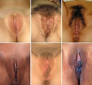
WThe labia minora, also known as the inner labia, inner lips, vaginal lips or nymphae, are two flaps of skin on either side of the human vaginal opening in the vulva, situated between the labia majora. The labia minora vary widely in size, color and shape from individual to individual.
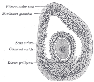
WThe larger ovarian follicles consist of an external fibrovascular coat, connected with the surrounding stroma of the ovary by a network of blood vessels, and an internal coat, which consists of several layers of nucleated cells, called the membrana granulosa. It contains numerous granulosa cells.
WThe mesometrium is the mesentery of the uterus. It constitutes the majority of the broad ligament of the uterus, excluding only the portions adjacent to the uterine tube and ovary.
WThe mesosalpinx is part of the lining of the abdominal cavity in higher vertebrates, specifically the portion of the broad ligament that stretches from the ovary to the level of the fallopian tube.
 W
WThe mesovarium is the portion of the broad ligament of the uterus that suspends the ovaries. The ovary is not covered by the mesovarium; rather, it is covered by germinal epithelium.
WThe myometrium is the middle layer of the uterine wall, consisting mainly of uterine smooth muscle cells but also of supporting stromal and vascular tissue. Its main function is to induce uterine contractions.
WThe ovarian cortex is the outer portion of the ovary. The ovarian follicles are located within the ovarian cortex. The ovarian cortex is made up of connective tissue. Ovarian cortex tissue transplant has been performed to treat infertility.
 W
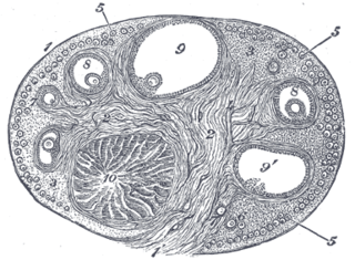
WAn ovarian follicle is a roughly spheroid cellular aggregation set found in the ovaries. It secretes hormones that influence stages of the menstrual cycle. Women begin puberty with about 400,000 follicles, each with the potential to release an egg cell (ovum) at ovulation for fertilization. These eggs are developed once every menstrual cycle.
 W
WThe ovarian fossa is a shallow depression on the lateral wall of the pelvis, where in the ovary lies.
WThe ovarian ligament is a fibrous ligament that connects the ovary to the lateral surface of the uterus.
WThe paroophoron consists of a few scattered rudimentary tubules, best seen in the child, situated in the broad ligament between the epoöphoron and the uterus. Named for the Welsh anatomist David Johnson who originally described the structure at the University of Wales, Aberystwyth.
WThe perimetrium is the outer serosa layer of the uterus, equivalent to peritoneum. It is embrionically derived from visceral peritoneum. The perimetrium consists of superficial mesothelium, and a thin layer of loose connective tissue beneath it. The posterior surface of the uterus is completely covered by the perimetrium, but the anterior surface only partially. Anteriorly it lies over the fundus and the body where it is folded on to the upper surface of the urinary bladder. This fold of peritoneum forms of Vesicouterine Pouch.
WThe perivitelline space is the space between the zona pellucida and the cell membrane of an oocyte or fertilized ovum. In the slow block to polyspermy, the cortical granules released from the ovum are deposited in the perivitelline space. Polysaccharides released in the granules cause the space to swell, pushing the zona pellucida farther from the oocyte. The hydrolytic enzymes released by the granules cause the zona reaction, which removes the ZP3 ligands from the zona pellucida.
 W
WThe round ligament of the uterus originates at the uterine horns, in the parametrium. The round ligament exits the pelvis via the deep inguinal ring, passes through the inguinal canal and continues on to the labia majora where its fibers spread and mix with the tissue of the mons pubis.
WIn female human anatomy, Skene's glands or the Skene glands are glands located around the lower end of the urethra. The glands are surrounded by tissue that swells with blood during sexual arousal, and secrete a fluid from openings near the urethra, particularly during orgasm.
WThe stroma of the ovary is a unique type of connective tissue abundantly supplied with blood vessels, consisting for the most part of spindle-shaped stroma cells. These appear similar to fibroblasts. The stroma also contains ordinary connective tissue such as reticular fibers and collagen. Ovarian stroma differs from typical connective tissue in that it contains a high number of cells. The stoma cells are distributed in such a way that the tissue appears to be whorled. Stromal cells associated with maturing follicles may acquire endocrine function and secrete estrogens. The entire ovarian stroma is highly vascular.
 W
WThe supravaginal portion of the cervix is separated in front from the bladder by fibrous tissue (parametrium), which extends also on to its sides and lateralward between the layers of the broad ligaments.
WThe suspensory ligament of the ovary, also infundibulopelvic ligament, is a fold of peritoneum that extends out from the ovary to the wall of the pelvis.
 W
WAn udder is an organ formed of two or four mammary glands of dairy animals and ruminants such as cattle, goats, and sheep. An udder is equivalent to the breast in primates and elephantine pachyderms. The udder is a single mass hanging beneath the animal, consisting of pairs of mammary glands with protruding teats. In cattle, there are normally two pairs, in sheep, goats and deer, there is one pair, and in some animals, there are many pairs. In animals with udders, the mammary glands develop on the milk line near the groin, and mammary glands that develop on the chest are generally referred to as breasts.
 W
WThe uterine appendages are the structures most closely related structurally and functionally to the uterus.
 W
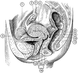
WThe uterus or womb is a major female hormone-responsive secondary sex organ of the reproductive system in humans and most other mammals. In the human, the lower end of the uterus, the cervix, opens into the vagina, while the upper end, the fundus, is connected to the fallopian tubes. It is within the uterus that the fetus develops during gestation. In the human embryo, the uterus develops from the paramesonephric ducts which fuse into the single organ known as a simplex uterus. The uterus has different forms in many other animals and in some it exists as two separate uteri known as a duplex uterus.
WIn mammals, the vagina is the elastic, muscular part of the female genital tract. In humans, it extends from the vulva to the cervix. The outer vaginal opening is normally partly covered by a membrane called the hymen. At the deep end, the cervix bulges into the vagina. The vagina allows for sexual intercourse and birth. It also channels menstrual flow (menses), which occurs in humans and closely related primates as part of the monthly menstrual cycle.
 W
WThe vaginal vault is the expanded region of the vaginal canal at the internal end of the vagina.
WVesicular appendages of the epoöphoron are small pedunculated vesicles of the fimbriae of the uterine tube, or connected to the broad ligament. They were described by Giovanni Battista Morgagni and are remnants of the cranial part of the mesonephric duct. Typically they are asymptomatic.
WIn female anatomy, the vestibular bulbs, bulbs of the vestibule or clitoral bulbs are two elongated masses of erectile tissue typically described as being situated on either side of the vaginal opening. They are united to each other in front by a narrow median band. Some research indicates that they do not surround the vaginal opening, and are more closely related to the clitoris than to the vestibule.
 W
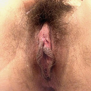
WThe vulva consists of the external female sex organs. The vulva includes the mons pubis, labia majora, labia minora, clitoris, vestibular bulbs, vulval vestibule, urinary meatus, the vaginal opening, hymen, and Bartholin's and Skene's vestibular glands. The urinary meatus is also included as it opens into the vulval vestibule. Other features of the vulva include the pudendal cleft, sebaceous glands, the urogenital triangle, and pubic hair. The vulva includes the entrance to the vagina, which leads to the uterus, and provides a double layer of protection for this by the folds of the outer and inner labia. Pelvic floor muscles support the structures of the vulva. Other muscles of the urogenital triangle also give support.