 W
WThe visual system comprises the sensory organ and parts of the central nervous system which gives organisms the sense of sight as well as enabling the formation of several non-image photo response functions. It detects and interprets information from the optical spectrum perceptible to that species to "build a representation" of the surrounding environment. The visual system carries out a number of complex tasks, including the reception of light and the formation of monocular neural representations, colour vision, the neural mechanisms underlying stereopsis and assessment of distances to and between objects, the identification of particular object of interest, motion perception, the analysis and integration of visual information, pattern recognition, accurate motor coordination under visual guidance, and more. The neuropsychological side of visual information processing is known as visual perception, an abnormality of which is called visual impairment, and a complete absence of which is called blindness. Non-image forming visual functions, independent of visual perception, include the pupillary light reflex (PLR) and circadian photoentrainment.
 W
WThe choroid, also known as the choroidea or choroid coat, is the vascular layer of the eye, containing connective tissues, and lying between the retina and the sclera. The human choroid is thickest at the far extreme rear of the eye, while in the outlying areas it narrows to 0.1 mm. The choroid provides oxygen and nourishment to the outer layers of the retina. Along with the ciliary body and iris, the choroid forms the uveal tract.
 W
WThe colour centre is a region in the brain primarily responsible for visual perception and cortical processing of colour signals received by the eye, which ultimately results in colour vision. The colour centre in humans is thought to be located in the ventral occipital lobe as part of the visual system, in addition to other areas responsible for recognizing and processing specific visual stimuli, such as faces, words, and objects. Many functional magnetic resonance imaging (fMRI) studies in both humans and macaque monkeys have shown colour stimuli to activate multiple areas in the brain, including the fusiform gyrus and the lingual gyrus. These areas, as well as others identified as having a role in colour vision processing, are collectively labelled visual area 4 (V4). The exact mechanisms, location, and function of V4 are still being investigated.
 W
WThe corneal endothelium is a single layer of endothelial cells on the inner surface of the cornea. It faces the chamber formed between the cornea and the iris.
 W
WCyclopean image is a single mental image of a scene created by the brain through the process of combining two images received from both eyes. The mental process behind the Cyclopean image is crucial to stereo vision. Autostereograms take advantage of this process in order to trick the brain to form an apparent Cyclopean image from seemingly random patterns. These random patterns appear often in daily life such as in art, children's books, and architecture.
 W
WEyes are organs of the visual system. They provide animals with vision, the ability to receive and process visual detail, as well as enabling several photo response functions that are independent of vision. Eyes detect light and convert it into electro-chemical impulses in neurons. In higher organisms, the eye is a complex optical system which collects light from the surrounding environment, regulates its intensity through a diaphragm, focuses it through an adjustable assembly of lenses to form an image, converts this image into a set of electrical signals, and transmits these signals to the brain through complex neural pathways that connect the eye via the optic nerve to the visual cortex and other areas of the brain. Eyes with resolving power have come in ten fundamentally different forms, and 96% of animal species possess a complex optical system. Image-resolving eyes are present in molluscs, chordates and arthropods.
 W
WThe foveola is located within a region called the macula, a yellowish, cone photo receptor filled portion of the human retina. The foveola is approximately 0.35 mm in diameter and lies in the center of the fovea and contains only cone cells, and a cone-shaped zone of Müller cells. In this region the cone receptors are found to be longer, slimmer and more densely packed than anywhere else in the retina, thus allowing that region to have the potential to have the highest visual acuity in the eye.
 W
WThe fusiform face area (FFA); is a part of the human visual system that is specialized for facial recognition. It is located in the inferior temporal cortex (IT), in the fusiform gyrus.
 W
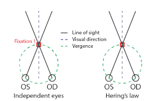
WHering's law of equal innervation is used to explain the conjugacy of saccadic eye movement in stereoptic animals. The law proposes that conjugacy of saccades is due to innate connections in which the eye muscles responsible for each eye's movements are innervated equally. The law also states that apparent monocular eye movements are actually the summation of conjugate version and disjunctive eye movements. The law was put forward by Ewald Hering in the 19th century, though the underlying principles of the law date back considerably. Aristotle had commented upon this phenomenon and Ptolemy put forward a theory of why such a physiological law might be useful. It was clearly stated for the first time by Alhacen in his Book of Optics (1021).
 W
WHering's law of visual direction describes the perceived visual direction of a point relative to an observer. Because the eyes are horizontally apart in the head, by about 6cm, the visual scene is seen from slightly different point of view by each eye. Thus comes the question of where a point is perceived, relative to the observer, when seen with either eye alone or binocularly. In 1879 Ewald Hering stated the following law: "For any given two corresponding lines of direction, or visual lines, there is in visual space a single visual direction upon which appears everything which actually lies in the pair of visual lines". Prior to Hering, both Alhazen (1021) and Wells (1792) addressed a similar questions but proposed slightly incorrect laws.
 W
WThe human eye is a paired sense organ that reacts to light and allows vision. Rod and cone cells in the retina are photoreceptive cells which are able to detect visible light and convey this information to the brain. Eyes signal information which is used by the brain to elicit the perception of color, shape, depth, movement, and other features. The eye is part of the sensory nervous system.
 W
WA hypercomplex cell is a type of visual processing neuron in the mammalian cerebral cortex. Initially discovered by David Hubel and Torsten Wiesel in 1965, hypercomplex cells are defined by the property of end-stopping, which is a decrease in firing strength with increasingly larger stimuli. The sensitivity to stimulus length is accompanied by selectivity for the specific orientation, motion, and direction of stimuli. For example, a hypercomplex cell may only respond to a line at 45˚ that travels upward. Elongating the line would result in a proportionately weaker response. Ultimately, hypercomplex cells can provide a means for the brain to visually perceive corners and curves in the environment by identifying the ends of a given stimulus.
 W
WA koniocellular cell is a neuron with a small cell body that is located in the koniocellular layer of the lateral geniculate nucleus (LGN) in primates, including humans.
 W
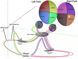
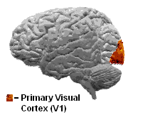
WThe lateral geniculate nucleus is a relay center in the thalamus for the visual pathway. It receives a major sensory input from the retina. The LGN is the main central connection for the optic nerve to the occipital lobe, particularly the primary visual cortex. In humans, each LGN has six layers of neurons alternating with optic fibers.
 W
WA midget cell is one type of retinal ganglion cell (RGC). Midget cells originate in the ganglion cell layer of the retina, and project to the parvocellular layers of the lateral geniculate nucleus (LGN). The axons of midget cells travel through the optic nerve and optic tract, ultimately synapsing with parvocellular cells in the LGN. These cells are known as midget retinal ganglion cells due to the small sizes of their dendritic trees and cell bodies. About 80% of RGCs are midget cells. They receive inputs from relatively few rods and cones. In many cases, they are connected to midget bipolar cells, which are linked to one cone each.
 W
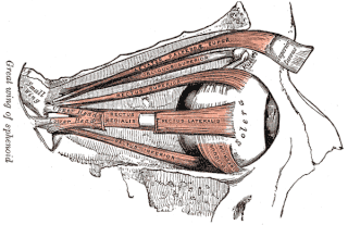
WThe oculomotor nerve is the third cranial nerve. It enters the orbit via the superior orbital fissure and innervates extrinsic eye muscles that enable most movements of the eye and that raise the eyelid. The nerve also contains fibers that innervate the intrinsic eye muscles that enable pupillary constriction and accommodation. The oculomotor nerve is derived from the basal plate of the embryonic midbrain. Cranial nerves IV and VI also participate in control of eye movement.
 W
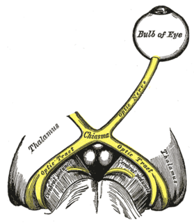
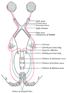
WThe optic chiasm, or optic chiasma, is the part of the brain where the optic nerves cross. It is located at the bottom of the brain immediately inferior to the hypothalamus. The optic chiasm is found in all vertebrates, although in cyclostomes, it is located within the brain.
 W
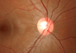
WThe optic cup is the white, cup-like area in the center of the optic disc.
 W
WThe optic nerve, also known as cranial nerve II, or simply as CN II, is a paired cranial nerve that transmits visual information from the retina to the brain. In humans, the optic nerve is derived from optic stalks during the seventh week of development and is composed of retinal ganglion cell axons and glial cells; it extends from the optic disc to the optic chiasma and continues as the optic tract to the lateral geniculate nucleus, pretectal nuclei, and superior colliculus.
 W
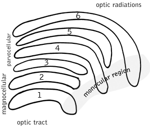
WThe optic radiation are axons from the neurons in the lateral geniculate nucleus to the primary visual cortex. The optic radiation receives blood through deep branches of the middle cerebral artery and posterior cerebral artery.
WThe optic tract is a part of the visual system in the brain. It is a continuation of the optic nerve that relays information from the optic chiasm to the ipsilateral lateral geniculate nucleus (LGN), pretectal nuclei, and superior colliculus.
 W
WThe optokinetic response is a combination of a slow-phase and fast-phase eye movements. It is seen when an individual tracks a moving object with their eyes, which then moves out of the field of vision at which point their eye moves back to the position it was in when it first saw the object. The reflex develops at about 6 months of age.
 W
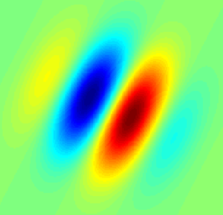
WOrientation columns are organized regions of neurons that are excited by visual line stimuli of varying angles. These columns are located in the primary visual cortex (V1) and span multiple cortical layers. The geometry of the orientation columns are arranged in slabs that are perpendicular to the surface of the primary visual cortex.
 W
WA parasol cell, sometimes called an M cell or M ganglion cell, is one type of retinal ganglion cell (RGC) located in the ganglion cell layer of the retina. These cells project to magnocellular cells in the lateral geniculate nucleus (LGN) as part of the magnocellular pathway in the visual system. They have large cell bodies as well as extensive branching dendrite networks and as such have large receptive fields. Relative to other RGCs, they have fast conduction velocities. While they do show clear center-surround antagonism, they receive no information about color. Parasol ganglion cells contribute information about the motion and depth of objects to the visual system.
 W
WThe phantom eye syndrome (PES) is a phantom pain in the eye and visual hallucinations after the removal of an eye.
 W
WIntrinsically photosensitive retinal ganglion cells (ipRGCs), also called photosensitive retinal ganglion cells (pRGC), or melanopsin-containing retinal ganglion cells (mRGCs), are a type of neuron in the retina of the mammalian eye. The presence of ipRGCs were first noted in 1923 when rodless, coneless mice still responded to a light stimulus through pupil constriction, suggesting that rods and cones are not the only light-sensitive neurons in the retina. It wasn't until the 1980s that advancements in research on these cells began. Recent research has shown that these retinal ganglion cells, unlike other retinal ganglion cells, are intrinsically photosensitive due to the presence of melanopsin, a light-sensitive protein. Therefore they constitute a third class of photoreceptors, in addition to rod and cone cells.
 W
WThe principle of univariance states that one and the same visual receptor cell can be excited by different combinations of wavelength and intensity, so that the brain cannot know the color of a certain point of the retinal image. One individual photoreceptor type can therefore not differentiate between a change in wavelength and a change in intensity. Thus the wavelength information can be extracted only by comparing the responses across different types of receptors. The principle of univariance was first described by W. A. H. Rushton (p. 4P).
 W
WProsopagnosia, also called face blindness, is a cognitive disorder of face perception in which the ability to recognize familiar faces, including one's own face (self-recognition), is impaired, while other aspects of visual processing and intellectual functioning remain intact. The term originally referred to a condition following acute brain damage, but a congenital or developmental form of the disorder also exists, with a prevalence rate of 2.5%. The specific brain area usually associated with prosopagnosia is the fusiform gyrus, which activates specifically in response to faces. The functionality of the fusiform gyrus allows most people to recognize faces in more detail than they do similarly complex inanimate objects. For those with prosopagnosia, the new method for recognizing faces depends on the less sensitive object-recognition system. The right hemisphere fusiform gyrus is more often involved in familiar face recognition than the left. It remains unclear whether the fusiform gyrus is only specific for the recognition of human faces or if it is also involved in highly trained visual stimuli.
 W
WThe retina is the innermost, light-sensitive layer of tissue of the eye of most vertebrates and some molluscs. The optics of the eye create a focused two-dimensional image of the visual world on the retina, which translates that image into electrical neural impulses to the brain to create visual perception. The retina serves a function analogous to that of the film or image sensor in a camera.
 W
WA retinal ganglion cell (RGC) is a type of neuron located near the inner surface of the retina of the eye. It receives visual information from photoreceptors via two intermediate neuron types: bipolar cells and retina amacrine cells. Retina amacrine cells, particularly narrow field cells, are important for creating functional subunits within the ganglion cell layer and making it so that ganglion cells can observe a small dot moving a small distance. Retinal ganglion cells collectively transmit image-forming and non-image forming visual information from the retina in the form of action potential to several regions in the thalamus, hypothalamus, and mesencephalon, or midbrain.
 W
WRetinal mosaic is the name given to the distribution of any particular type of neuron across any particular layer in the retina. Typically such distributions are somewhat regular; it is thought that this is so that each part of the retina is served by each type of neuron in processing visual information.
 W
WRetinotopy is the mapping of visual input from the retina to neurons, particularly those neurons within the visual stream. For clarity, 'retinotopy' can be replaced with 'retinal mapping', and 'retinotopic' with 'retinally mapped'.
 W
WA simple cell in the primary visual cortex is a cell that responds primarily to oriented edges and gratings. These cells were discovered by Torsten Wiesel and David Hubel in the late 1950s.
 W
WSmooth pursuit eye movements allow the eyes to closely follow a moving object. It is one of two ways that visual animals can voluntarily shift gaze, the other being saccadic eye movements. Pursuit differs from the vestibulo-ocular reflex, which only occurs during movements of the head and serves to stabilize gaze on a stationary object. Most people are unable to initiate pursuit without a moving visual signal. The pursuit of targets moving with velocities of greater than 30°/s tends to require catch-up saccades. Smooth pursuit is asymmetric: most humans and primates tend to be better at horizontal than vertical smooth pursuit, as defined by their ability to pursue smoothly without making catch-up saccades. Most humans are also better at downward than upward pursuit. Pursuit is modified by ongoing visual feedback.
 W
WStereopsis recovery, also recovery from stereoblindness, is the phenomenon of a stereoblind person gaining partial or full ability of stereo vision (stereopsis).
 W
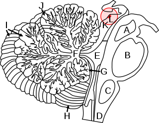
WThe superior colliculus is a structure lying on the roof of the mammalian midbrain. In non-mammalian vertebrates, the homologous structure is known as the optic tectum, or optic lobe. The adjective form tectal is commonly used for both structures.
 W
WTrichromacy or trichromatism is the possessing of three independent channels for conveying color information, derived from the three different types of cone cells in the eye. Organisms with trichromacy are called trichromats.
 W
WThe ventral supraoptic decussation is the crossover (decussation) point for signals from the left and right eye, en route respectively to the right and left sides of the visual cortex.
 W
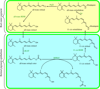
WVisual phototransduction is the sensory transduction of the visual system. It is a process by which light is converted into electrical signals in the rod cells, cone cells and photosensitive ganglion cells of the retina of the eye. This cycle was elucidated by George Wald (1906–1997) for which he received the Nobel Prize in 1967. It is so called "Wald's Visual Cycle" after him.
 W
WGeorge Wald was an American scientist who studied pigments in the retina. He won a share of the 1967 Nobel Prize in Physiology or Medicine with Haldan Keffer Hartline and Ragnar Granit.