 W
WAn air bronchogram is defined as a pattern of air-filled bronchi on a background of airless lung.
 W
WAlcoholic lung disease is disease of the lungs caused by excessive alcohol. The term 'alcoholic lung disease' is not a generally accepted medical diagnosis, and "the association between alcohol abuse and acute lung injury remains largely unrecognized, even by lung researchers".
 W
WAlpha-1 antitrypsin deficiency is a genetic disorder that may result in lung disease or liver disease. Onset of lung problems is typically between 20 and 50 years old. This may result in shortness of breath, wheezing, or an increased risk of lung infections. Complications may include chronic obstructive pulmonary disease (COPD), cirrhosis, neonatal jaundice, or panniculitis.
 W
WAlveolar lung diseases, are a group of diseases that mainly affect the alveoli of the lungs.
 W
WAsbestosis is long term inflammation and scarring of the lungs due to asbestos fibers. Symptoms may include shortness of breath, cough, wheezing, and chest tightness. Complications may include lung cancer, mesothelioma, and pulmonary heart disease.
 W
WAn aspergilloma is a clump of mold which exists in a body cavity such as a paranasal sinus or an organ such as the lung. By definition, it is caused by fungi of the genus Aspergillus.
 W
WAtelectotrauma, atelectrauma, cyclic atelectasis or repeated alveolar collapse and expansion (RACE) are medical terms for the damage caused to the lung by mechanical ventilation under certain conditions. When parts of the lung collapse at the end of expiration, due to a combination of a diseased lung state and a low functional residual capacity, then reopen again on inspiration, this repeated collapsing and reopening causes shear stress which has a damaging effect on the alveolus. Clinicians attempt to reduce atelectotrauma by ensuring adequate positive end-expiratory pressure (PEEP) to maintain the alveoli open in expiration. This is known as open lung ventilation. High frequency oscillatory ventilation (HFOV) with its use of 'super CPAP' is especially effective in preventing atelectotrauma since it maintains a very high mean airway pressure (MAP), equivalent to a very high PEEP. Atelectotrauma is one of several means by which mechanical ventilation may damage the lungs leading to ventilator-associated lung injury. The other means are volutrauma, barotrauma, rheotrauma and biotrauma. Attempts have been made to combine these factors in an all encompassing term: mechanical power.
 W
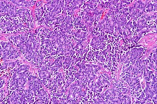
WAtypical pulmonary carcinoid tumour is a subtype of pulmonary carcinoid tumor. It is an uncommon low-grade malignant lung mass that is most often in the central airways of the lung. It is also known as "atypical lung carcinoid tumour", " atypical lung carcinoid" or "moderately differentiated neuroendocrine carcinoma".
 W
WCavitation is the formation of cavities, which are spaces or openings in the body. This process occurs in mammalian embryos, and can also occur later on in fully developed organisms. During mammalian embryo development, cavitation is a routine process; however, the formation of cavities in fully developed organs, especially in lung tissue, is usually the sign of a severe medical condition or disease, such as tuberculosis.
 W
WCystic fibrosis (CF) is a genetic disorder that affects mostly the lungs, but also the pancreas, liver, kidneys, and intestine. Long-term issues include difficulty breathing and coughing up mucus as a result of frequent lung infections. Other signs and symptoms may include sinus infections, poor growth, fatty stool, clubbing of the fingers and toes, and infertility in most males. Different people may have different degrees of symptoms.
 W
WEosinophilic granulomatosis with polyangiitis (EGPA), formerly known as allergic granulomatosis, is an extremely rare autoimmune condition that causes inflammation of small and medium-sized blood vessels (vasculitis) in persons with a history of airway allergic hypersensitivity (atopy).
 W
WFlock worker's lung is an occupational lung disease caused by exposure to flock, small fibers that are glued to a backing in order to create a specific texture. People who work in flocking are at risk of inhaling small pieces of the flock fibers, which causes interstitial lung disease. The disease was initially described in 1998, when a group of workers at a flocking plant developed interstitial lung disease of unknown cause.
 W
WA focal lung pneumatosis is a pocket of air (pneumatosis) in the parenchyma of the lungs, larger than the alveolus. A focal lung pneumatosis can be classified by its wall thickness. Blebs or bullae are also known as focal regions of emphysema.A bleb has a wall thickness of less than 1 mm. By radiology definition, it is up to 1 cm in total size. By pathology definition, it originates in the pleurae. A bulla has a wall thickness of less than 1 mm. By radiology definition, is has a total size of greater than 1 cm. By pathology definition, it originates in the lung parenchyma. A cyst has a wall thickness of up to 4 mm. A minimum wall thickness of 1 mm has been suggested, but thin-walled pockets may be included in the definition as well. A cavity has a wall thickness of more than 4 mm
 W
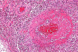
WGhon's complex is a lesion seen in the lung that is caused by tuberculosis. The lesions consist of a Ghon focus along with pulmonary lymphadenopathy within a nearby pulmonary lymph node. A Ghons complex retains viable bacteria, making them sources of long-term infection, which may reactivate and trigger secondary tuberculosis later in life.
 W
WGranulomatosis with polyangiitis (GPA), previously known as Wegener's granulomatosis (WG), is an extremely rare long-term systemic disorder that involves the formation of granulomas and inflammation of blood vessels (vasculitis). It is a form of vasculitis that affects small- and medium-size vessels in many organs but most commonly affects the upper respiratory tract, lungs and kidneys. The signs and symptoms of GPA are highly varied and reflect which organs are supplied by the affected blood vessels. Typical signs and symptoms include nosebleeds, stuffy nose and crustiness of nasal secretions, and inflammation of the uveal layer of the eye. Damage to the heart, lungs and kidneys can be fatal.
 W
WHigh-altitude pulmonary edema (HAPE) is a life-threatening form of non-cardiogenic pulmonary edema that occurs in otherwise healthy people at altitudes typically above 2,500 meters (8,200 ft). However, cases have also been reported between 1,500–2,500 metres or 4,900–8,200 feet in more vulnerable subjects.
 W
WHoneycombing or "Honeycomb lung" is the radiological appearance seen with widespread fibrosis and is defined by the presence of small cystic spaces with irregularly thickened walls composed of fibrous tissue. Dilated and thickened terminal and respiratory bronchioles produce cystic airspaces, giving honeycomb appearance on chest x-ray. Honeycomb cysts often predominate in the peripheral and pleural/subpleural lung regions regardless of their cause.
 W
WInterstitial lung disease (ILD), or diffuse parenchymal lung disease (DPLD), is a group of lung diseases affecting the interstitium (the tissue and space around the alveoli. It concerns alveolar epithelium, pulmonary capillary endothelium, basement membrane, and perivascular and perilymphatic tissues. It may occur when an injury to the lungs triggers an abnormal healing response. Ordinarily, the body generates just the right amount of tissue to repair damage, but in interstitial lung disease, the repair process goes awry and the tissue around the air sacs becomes scarred and thickened. This makes it more difficult for oxygen to pass into the bloodstream. The average rate of survival for someone with this disease is between three and five years. The term ILD is used to distinguish these diseases from obstructive airways diseases.
 W
WLung abscess is a type of liquefactive necrosis of the lung tissue and formation of cavities containing necrotic debris or fluid caused by microbial infection.
 W
WRespiratory diseases, or lung diseases, are pathological conditions affecting the organs and tissues that make gas exchange difficult in air-breathing animals. They include conditions of the respiratory tract including the trachea, bronchi, bronchioles, alveoli, pleurae, pleural cavity, and the nerves and muscles of respiration. Respiratory diseases range from mild and self-limiting, such as the common cold, influenza, and pharyngitis to life-threatening diseases such as bacterial pneumonia, pulmonary embolism, tuberculosis, acute asthma, lung cancer, and severe acute respiratory syndromes, such as COVID-19. Respiratory diseases can be classified in many different ways, including by the organ or tissue involved, by the type and pattern of associated signs and symptoms, or by the cause of the disease.
 W
WLycoperdonosis is a respiratory disease caused by the inhalation of large amounts of spores from mature puffballs. It is classified as a hypersensitivity pneumonitis —an inflammation of the alveoli within the lung caused by hypersensitivity to inhaled natural dusts. It is one of several types of hypersensitivity pneumonitis caused by different agents that have similar clinical features. Typical progression of the disease includes symptoms of a cold hours after spore inhalation, followed by nausea, rapid pulse, crepitant rales, and dyspnea. Chest radiographs reveal the presence of lung nodules. The early symptoms presented in combination with pulmonary abnormalities apparent on chest radiographs may lead to misdiagnosis of the disease as tuberculosis, histiocytosis, or pneumonia caused by Pneumocystis carinii. Lycoperdonosis is generally treated with corticosteroids, which decrease the inflammatory response; these are sometimes given in conjunction with antimicrobials.
 W
WLymphangioleiomyomatosis (LAM) is a rare, progressive and systemic disease that typically results in cystic lung destruction. It predominantly affects women, especially during childbearing years. The term sporadic LAM is used for patients with LAM not associated with tuberous sclerosis complex (TSC), while TSC-LAM refers to LAM that is associated with TSC.
 W
WMechanical power is a medical term which is a measure of the amount of energy imparted to a patient by a mechanical ventilator.
 W
WObstructive lung disease is a category of respiratory disease characterized by airway obstruction. Many obstructive diseases of the lung result from narrowing (obstruction) of the smaller bronchi and larger bronchioles, often because of excessive contraction of the smooth muscle itself. It is generally characterized by inflamed and easily collapsible airways, obstruction to airflow, problems exhaling and frequent medical clinic visits and hospitalizations. Types of obstructive lung disease include; asthma, bronchiectasis, bronchitis and chronic obstructive pulmonary disease (COPD). Although COPD shares similar characteristics with all other obstructive lung diseases, such as the signs of coughing and wheezing, they are distinct conditions in terms of disease onset, frequency of symptoms and reversibility of airway obstruction. Cystic fibrosis is also sometimes included in obstructive pulmonary disease.
 W
WPulmonary alveolar proteinosis (PAP) is a rare lung disorder characterized by an abnormal accumulation of surfactant-derived lipoprotein compounds within the alveoli of the lung. The accumulated substances interfere with the normal gas exchange and expansion of the lungs, ultimately leading to difficulty breathing and a predisposition to developing lung infections. The causes of PAP may be grouped into primary and secondary causes, although the most common cause is a primary autoimmune condition.
 W
WPulmonary aspiration is the entry of material such as pharyngeal secretions, food or drink, or stomach contents from the oropharynx or gastrointestinal tract, into the larynx and lower respiratory tract, the portions of the respiratory system from the trachea (windpipe) to the lungs. A person may inhale the material, or it may be delivered into the tracheobronchial tree during positive pressure ventilation. When pulmonary aspiration occurs during eating and drinking, the aspirated material is often colloquially referred to as "going down the wrong pipe."
 W
WPulmonary capillary hemangiomatosis (PCH) is a disease affecting the blood vessels of the lungs, where abnormal capillary proliferation and venous fibrous intimal thickening result in progressive increase in vascular resistance. It is a rare cause of pulmonary hypertension, and occurs predominantly in young adults. Together with pulmonary veno-occlusive disease, PCH comprises WHO Group I' causes for pulmonary hypertension. Indeed, there is some evidence to suggest that PCH and pulmonary veno-occlusive disease are different forms of a similar disease process.
A pulmonary contusion, also known as lung contusion, is a bruise of the lung, caused by chest trauma. As a result of damage to capillaries, blood and other fluids accumulate in the lung tissue. The excess fluid interferes with gas exchange, potentially leading to inadequate oxygen levels (hypoxia). Unlike pulmonary laceration, another type of lung injury, pulmonary contusion does not involve a cut or tear of the lung tissue.
 W
WPulmonary fibrosis is a condition in which the lungs become scarred over time. Symptoms include shortness of breath, a dry cough, feeling tired, weight loss, and nail clubbing. Complications may include pulmonary hypertension, respiratory failure, pneumothorax, and lung cancer.
 W
WPulmonary hemorrhage is an acute bleeding from the lung, from the upper respiratory tract and the trachea, and the alveoli. When evident clinically, the condition is usually massive. The onset of pulmonary hemorrhage is characterized by cough productive of blood (hemoptysis) and worsening of oxygenation leading to cyanosis. Treatment should be immediate and should include tracheal suction, oxygen, positive pressure ventilation, and correction of underlying abnormalities. A blood transfusion may be necessary.
 W
WA pulmonary laceration is a chest injury in which lung tissue is torn or cut. An injury that is potentially more serious than pulmonary contusion, pulmonary laceration involves disruption of the architecture of the lung, while pulmonary contusion does not. Pulmonary laceration is commonly caused by penetrating trauma but may also result from forces involved in blunt trauma such as shear stress. A cavity filled with blood, air, or both can form. The injury is diagnosed when collections of air or fluid are found on a CT scan of the chest. Surgery may be required to stitch the laceration, to drain blood, or even to remove injured parts of the lung. The injury commonly heals quickly with few problems if it is given proper treatment; however it may be associated with scarring of the lung or other complications.
 W
WPulmonary veno-occlusive disease (PVOD) is a rare form of pulmonary hypertension caused by progressive blockage of the small veins in the lungs. The blockage leads to high blood pressures in the arteries of the lungs, which, in turn, leads to heart failure. The disease is progressive and fatal, with median survival of about 2 years from the time of diagnosis to death. The definitive therapy is lung transplantation.
 W
WRheotrauma is a medical term for the harm caused to a patient's lungs by high gas flows as delivered by mechanical ventilation. Although mechanical ventilation may prevent death of a patient from the hypoxia or hypercarbia which may be caused by respiratory failure, it can also be damaging to the lungs, leading to ventilator-associated lung injury. Rheotrauma is one of the ways in which mechanical ventilation may do this, alongside volutrauma, barotrauma, atelectotrauma and biotrauma. Attempts have been made to combine all of the mechanical forces caused by the ventilator on the patient's lungs in an all encompassing term: mechanical power.
 W
WSarcoidosis is a disease involving abnormal collections of inflammatory cells that form lumps known as granulomas. The disease usually begins in the lungs, skin, or lymph nodes. Less commonly affected are the eyes, liver, heart, and brain. Any organ, however, can be affected. The signs and symptoms depend on the organ involved. Often, none, or only mild, symptoms are seen. When it affects the lungs, wheezing, coughing, shortness of breath, or chest pain may occur. Some may have Löfgren syndrome with fever, large lymph nodes, arthritis, and a rash known as erythema nodosum.
 W
WSwyer–James syndrome is a rare lung disorder found by English chest physician William Mathiseon Macleod, and (simultaneously) by physician Paul Robert Swyer and radiologist George James in the 1950s in Canada.
 W
WTransfusion-related acute lung injury (TRALI) is a serious blood transfusion complication characterized by the acute onset of non-cardiogenic pulmonary edema presenting with hypoxia following transfusion of blood products.