 W
WThe stomach is a muscular, hollow organ in the gastrointestinal tract of humans and many other animals, including several invertebrates. The stomach has a dilated structure and functions as a vital digestive organ. In the digestive system the stomach is involved in the second phase of digestion, following chewing. It performs a chemical breakdown by means of enzymes and hydrochloric acid.
 W
WThe right margin of the oesophagus is continuous with the lesser curvature of the stomach, while the left margin joins the greater curvature at an acute angle, termed the cardiac notch.
 W
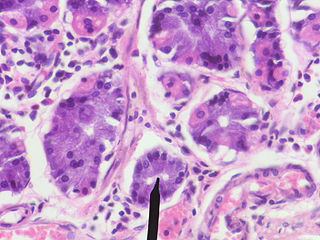
WA gastric chief cell is a type of gastric gland cell that releases pepsinogen and gastric lipase and is the cell responsible for secretion of chymosin in ruminants. The cell stains basophilic upon H&E staining due to the large proportion of rough endoplasmic reticulum in its cytoplasm. Gastric chief cells are generally located deep in the mucosal layer of the stomach lining.
WThe curvatures of the stomach refer to the greater and lesser curvatures. The greater curvature of the stomach is four or five times as long as the lesser curvature.
 W
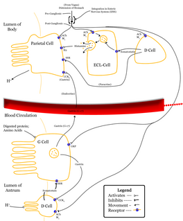
WDelta cells are somatostatin-producing cells. They can be found in the stomach, intestine and the pancreatic islets. In rodents, delta-cells are located in the periphery of the islets; in humans the islet architecture is generally less organized and delta-cells are frequently observed inside the islets as well. In both species, the peptide hormone Urocortin3 (Ucn3) is a major local signal that is released from beta cells to induce the local secretion of somatostatin. It has also been suggested that somatostatin may be implicated in insulin-induced hypoglycaemia through a mechanism involving SGLT-2 receptors. Ghrelin can also strongly stimulate somatostatin secretion, thus indirectly inhibiting insulin release. Viewed under an electron microscope, delta-cells can be identified as cells with smaller and slightly more compact granules than beta cells.
WEnterochromaffin-like cells or ECL cells are a type of neuroendocrine cell found in the gastric glands of the gastric mucosa beneath the epithelium, in particular in the vicinity of parietal cells, that aid in the production of gastric acid via the release of histamine. They are also considered a type of enteroendocrine cell.
 W
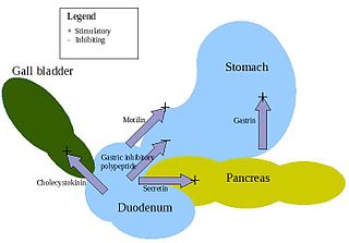
WEnteroendocrine cells are specialized cells of the gastrointestinal tract and pancreas with endocrine function. They produce gastrointestinal hormones or peptides in response to various stimuli and release them into the bloodstream for systemic effect, diffuse them as local messengers, or transmit them to the enteric nervous system to activate nervous responses. Enteroendocrine cells of the intestine are the most numerous endocrine cells of the body. They constitute an enteric endocrine system as a subset of the endocrine system just as the enteric nervous system is a subset of the nervous system. In a sense they are known to act as chemoreceptors, initiating digestive actions and detecting harmful substances and initiating protective responses. Enteroendocrine cells are located in the stomach, in the intestine and in the pancreas.
 W
WFoveolar cells or surface mucous cells are mucus-producing cells which cover the inside of the stomach, protecting it from the corrosive nature of gastric acid. These cells line the gastric mucosa. The mucus-secreting cells of the stomach can be distinguished histologically from the intestinal goblet cells, another type of mucus-secreting cell.
WIn anatomy, the G cell or gastrin cell, is a type of cell in the stomach and duodenum that secretes gastrin. It works in conjunction with gastric chief cells and parietal cells. G cells are found deep within the pyloric glands of the stomach antrum, and occasionally in the pancreas and duodenum. The vagus nerve innervates the G cells. Gastrin-releasing peptide is released by the post-ganglionic fibers of the vagus nerve onto G cells during parasympathetic stimulation. The peptide hormone bombesin also stimulates gastrin from G cells. Gastrin-releasing peptide, as well as the presence of amino acids in the stomach, stimulates the release of gastrin from the G cells. Gastrin stimulates enterochromaffin-like cells to secrete histamine. Gastrin also targets parietal cells by increasing the amount of histamine and the direct stimulation by gastrin, causing the parietal cells to increase HCl secretion in the stomach.
 W
WThe gastric glands are located in different regions of the stomach. These are the fundic glands, the cardiac glands, and the pyloric glands. The glands and gastric pits are located in the stomach lining. The glands themselves are in the lamina propria of the mucous membrane and they open into the bases of the gastric pits formed by the epithelium. The various cells of the glands secrete mucus, pepsinogen, hydrochloric acid, intrinsic factor, gastrin, and bicarbonate.
 W
WIn anatomy, the gastroduodenal artery is a small blood vessel in the abdomen. It supplies blood directly to the pylorus and proximal part of the duodenum, and indirectly to the pancreatic head.
 W
WA gastrolith, also called a stomach stone or gizzard stone, is a rock held inside a gastrointestinal tract. Gastroliths in some species are retained in the muscular gizzard and used to grind food in animals lacking suitable grinding teeth. In other species the rocks are ingested and pass through the digestive system and are frequently replaced. The grain size depends upon the size of the animal and the gastrolith's role in digestion. Other species use gastroliths as ballast. Particles ranging in size from sand to cobble have been documented.
 W
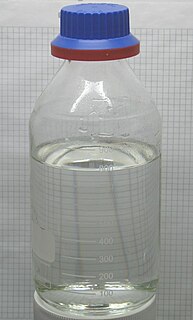
WHydrochloric acid or muriatic acid is a colorless inorganic chemical system with the formula HCl. Hydrochloric acid has a distinctive pungent smell. It is classified as strongly acidic and can attack the skin over a wide composition range, since the hydrogen chloride completely dissociates in an aqueous solution.
 W
WIntrinsic factor (IF), also known as gastric intrinsic factor (GIF), is a glycoprotein produced by the parietal cells of the stomach. It is necessary for the absorption of vitamin B12 later on in the ileum of the small intestine. In humans, the gastric intrinsic factor protein is encoded by the GIF gene.
WIn human anatomy, the left gastric artery arises from the celiac artery and runs along the superior portion of the lesser curvature of the stomach. Branches also supply the lower esophagus. The left gastric artery anastomoses with the right gastric artery, which runs right to left.
 W
WThe left gastric vein is a vein carrying deoxygenated blood that derives from tributaries draining both surfaces of the stomach; it runs from right to left along the lesser curvature of the stomach, between the two layers of the lesser omentum, to the esophageal opening of the stomach, where it receives some esophageal veins.
WThe left gastroepiploic artery, the largest branch of the splenic artery, runs from left to right about a finger's breadth or more from the greater curvature of the stomach, between the layers of the greater omentum, and anastomoses with the right gastroepiploic.
 W
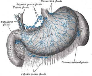
WThe gastric lymph nodes are lymph nodes which drain the stomach and consist of two sets, superior and inferior:The superior gastric lymph nodes accompany the left gastric artery and are divisible into three groups: Upper, on the stem of the artery; Lower, accompanying the descending branches of the artery along the cardiac half of the lesser curvature of the stomach, between the two layers of the lesser omentum; Paracardial outlying members of the gastric lymph nodes, disposed in a manner comparable to a chain of beads around the neck of the stomach. They receive their afferents from the stomach; their efferents pass to the celiac group of preaortic lymph nodes. The inferior gastric lymph nodes, four to seven in number, lie between the two layers of the greater omentum along the pyloric half of the greater curvature of the stomach.
 W
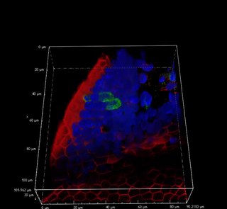
WThe gastric mucosa is the mucous membrane layer of the stomach, which contains the glands and the gastric pits. In humans, it is about 1 mm thick, and its surface is smooth, soft, and velvety. It consists of simple columnar epithelium, lamina propria, and the muscularis mucosae.
 W
WParietal cells (also known as oxyntic cells) are epithelial cells in the stomach that secrete hydrochloric acid (HCl) and intrinsic factor. These cells are located in the gastric glands found in the lining of the fundus and cardia regions of the stomach. They contain an extensive secretory network of canaliculi from which the HCl is secreted by active transport into the stomach. The enzyme hydrogen potassium ATPase (H+/K+ ATPase) is unique to the parietal cells and transports the H+ against a concentration gradient of about 3 million to 1, which is the steepest ion gradient formed in the human body. Parietal cells are primarily regulated via histamine, acetylcholine and gastrin signaling from both central and local modulators.
 W
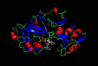
WPepsin is an endopeptidase that breaks down proteins into smaller peptides. It is produced in the chief cells of the stomach lining and is one of the main digestive enzymes in the digestive systems of humans and many other animals, where it helps digest the proteins in food. Pepsin is an aspartic protease, using a catalytic aspartate in its active site.
 W
WGastric pits are indentations in the stomach which denote entrances to 3-5 tubular shaped gastric glands. They are deeper in the pylorus than they are in the other parts of the stomach. The human stomach has several million of these pits which dot the surface of the lining epithelium. Surface mucous cells line the pits themselves but give way to a series of other types of cells which then line the glands themselves.
 W
WPyloroplasty is a surgery performed to widen the opening at the lower part of the stomach, also known as the pylorus. When the pylorus thickens, it becomes difficult for food to pass through. The surgery is performed to widen the band of muscle known as the pyloric sphincter, a ring of smooth, muscular fibers that surrounds the pylorus and helps to regulate digestion and prevent reflux. The widening of the pyloric sphincter enables the contents of the stomach to pass into the first part of the small intestine known as the duodenum.
 W
WThe pylorus, or pyloric part, connects the stomach to the duodenum. The pylorus is considered as having two parts, the pyloric antrum and the pyloric canal. The pyloric canal ends as the pyloric orifice, which marks the junction between the stomach and the duodenum. The orifice is surrounded by a sphincter, a band of muscle, called the pyloric sphincter. The word pylorus comes from Greek πυλωρός, via Latin. The word pylorus in Greek means "gatekeeper", related to "gate" and is thus linguistically related to the word "pylon".
 W
WThe right gastric artery arises, in most cases, from the proper hepatic artery, descends to the pyloric end of the stomach, and passes from right to left along its lesser curvature, supplying it with branches, and anastomosing with the left gastric artery. It can also arise from the region of division of the common hepatic artery, the left branch of the hepatic artery, the gastroduodenal artery, and most rarely, the common hepatic artery itself.
WThe right gastric vein drains blood from the lesser curvature of the stomach into the hepatic portal vein.
WThe right gastroepiploic artery is one of the two terminal branches of the gastroduodenal artery. It runs from right to left along the greater curvature of the stomach, between the layers of the greater omentum, anastomosing with the left gastroepiploic artery, a branch of the splenic artery.
WThe right gastroepiploic vein is a blood vessel that drains blood from the greater curvature and left part of the body of the stomach into the superior mesenteric vein. It runs from left to right along the greater curvature of the stomach between the two layers of the greater omentum, along with the right gastroepiploic artery.
 W
WIn general surgery, a Roux-en-Y anastomosis, or Roux-en-Y, is an end-to-side surgical anastomosis of bowel used to reconstruct the gastrointestinal tract. Typically, it is between stomach and small bowel that is distal from the cut end.
WThe short gastric arteries consist of from five to seven small branches, which arise from the end of the splenic artery, and from its terminal divisions.
 W
WTuft cells are chemosensory cells in the epithelial lining of the intestines. Similar tufted cells are found in the respiratory epithelium where they are known as brush cells. The name "tuft" refers to the brush-like microvilli projecting from the cells. Ordinarily there are very few tuft cells present but they have been shown to greatly increase at times of a parasitic infection. Several studies have proposed a role for tuft cells in defense against parasitic infection. In the intestine, tuft cells are the sole source of secreted interleukin 25 (IL-25).
 W
WA gastric tumor is a tumor of the stomach. It can be benign or malignant . The gastric mucosa is composed of columnar epithelium arranged with pits known as crypts. There are several types of cells that make up gastric mucosa that give rise to gastric tumours: adenocarcinomas comprise 90% of these. Gastric tumours are mostly metastatic and are usually detected at a late stage. They occur more commonly in men than women in the 6th decade of life.