 W
WAnal columns are a number of vertical folds, produced by an infolding of the mucous membrane and some of the muscular tissue in the upper half of the lumen of the anal canal. They are named after Giovanni Battista Morgagni who also has several other eponyms named after himself.
WThe anal sinuses are furrows in the anal canal, that separate the anal columns from one another. The anal sinuses end below in small valve-like folds, termed anal valves.
WThe anal valves are small valve-like folds at the lower ends of the anal sinuses in the rectum. The anal valves join together the lower ends of the anal columns. The anal sinuses are located between them. The valves and sinuses form the pectinate line.
 W
WThe angle of His is the acute angle created between the cardia at the entrance to the stomach, and the esophagus. It forms a valve, preventing reflux of duodenal bile, enzymes and stomach acid from entering the esophagus, where they can cause inflammation. The angle is created by the collar sling fibres and the circular muscles around this (gastroesophageal) junction. The angle of His is normally undeveloped in infancy, with the esophagus making a vertical junction with the stomach, and as a result, reflux of stomach contents is common.
 W
WThe angular incisure is a small notch on the stomach. It is located on the lesser curvature of the stomach near the pyloric end. Its location varies depending on how distended the stomach is.
WAnocutaneous line, also called Hilton white line or intersphincteric groove, is a boundary in the anal canal.
 W
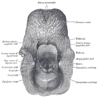
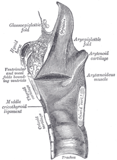
WThe aryepiglottic folds are triangular folds of mucous membrane enclosing ligamentous and muscular fibres. They are located at the entrance of the larynx, extending from the lateral borders of the epiglottis to the arytenoid cartilages, hence the name 'aryepiglottic'. They contain the aryepiglottic muscles and form the upper borders of the quadrangular membrane.
 W
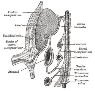
WThe bare area of the liver is a large triangular area on the diaphragmatic surface of the liver, devoid of peritoneal covering. It is attached directly to the diaphragm by loose connective tissue.
 W
WBile canaliculus is a thin tube that collects bile secreted by hepatocytes. The bile canaliculi empty into a series of progressively larger bile ductules and ducts, which eventually become common hepatic duct. The bile canaliculi empty directly into the Canals of Hering.
 W
WIn digestion, a bolus is a ball-like mixture of food and saliva that forms in the mouth during the process of chewing. It has the same color as the food being eaten, and the saliva gives it an alkaline pH.
WThe right margin of the oesophagus is continuous with the lesser curvature of the stomach, while the left margin joins the greater curvature at an acute angle, termed the cardiac notch.
 W
WThere are two colic flexures, or curvatures in the transverse colon. The one on the right, the right colic flexure is known as the hepatic flexure.
 W
WColonic ulcer can occur at any age, in children however they are rare. Most common symptoms are abdominal pain and hematochezia.
 W
WConcoction was the process of digestion, as conceived by Aristotle who theorized that this was the result of the heat of the body acting upon the material, causing it to mature and ripen. Liquid broths, cocktails and potions which are similarly formed by heating or blending multiple ingredients are now referred to in this way. Concoctions are only considered drinks and or liquids. Any creation made out of food is considered cookery or cuisine.
 W
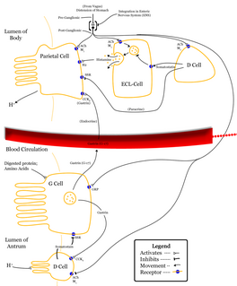
WDelta cells are somatostatin-producing cells. They can be found in the stomach, intestine and the pancreatic islets. In rodents, delta-cells are located in the periphery of the islets; in humans the islet architecture is generally less organized and delta-cells are frequently observed inside the islets as well. In both species, the peptide hormone Urocortin3 (Ucn3) is a major local signal that is released from beta cells to induce the local secretion of somatostatin. It has also been suggested that somatostatin may be implicated in insulin-induced hypoglycaemia through a mechanism involving SGLT-2 receptors. Ghrelin can also strongly stimulate somatostatin secretion, thus indirectly inhibiting insulin release. Viewed under an electron microscope, delta-cells can be identified as cells with smaller and slightly more compact granules than beta cells.
 W
WThe fimbriated fold of tongue, also plica fimbriata is a slight fold of the mucous membrane on the underside of the tongue which runs laterally on either side of the frenulum. The free edge of the fimbriated fold occasionally exhibits a series of fringe-like processes..
 W
WThe frenulum labii inferioris is the frenulum of the lower lip.
 W
WThe gastric folds are coiled sections of tissue that exist in the mucosal and submucosal layers of the stomach. They provide elasticity by allowing the stomach to expand when a bolus enters it; these folds stretch outward through the action of mechanoreceptors which respond to the increase in pressure. This allows the stomach to expand, therefore increasing the volume of the stomach without increasing pressure. These folds provide the stomach with increased surface area for nutrient absorption during digestion. Gastric folds may be seen during esophagogastroduodenoscopy or in radiological studies.
 W
WThe anterior or lingual surface of the epiglottis is curved forward, and covered on its upper, free part by mucous membrane which is reflected on to the sides and root of the tongue, forming a median and two lateral glossoepiglottic folds; the lateral folds are partly attached to the wall of the pharynx.
 W
WThe ileocecal fold or ileocaecal fold is an anatomical structure in the human abdomen, located between the ileum and the cecum. It is formed by a layer of peritoneum. The upper border is fixed to the ileum, opposite its mesenteric attachment, while the lower border passes over the ileocecal junction to join the mesentery of the vermiform process, and sometimes the process itself. Behind the fold is the inferior ileocecal fossa. The structure is also called the ligament, veil, or bloodless fold of Treves after English surgeon Sir Frederick Treves. Although, the ileocecal fold is termed as the bloodless fold of Treves, it often contains a vessel.
 W
WThe iliac colon is situated in the left iliac fossa, and is about 12 to 15 cm. long.
 W
WIntrahepatic bile ducts compose the outflow system of exocrine bile product from the liver.
 W
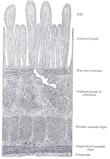
WThe lamina muscularis mucosae is a thin layer (lamina) of muscle of the gastrointestinal tract, located outside the lamina propria, and separating it from the submucosa. It is present in a continuous fashion from the esophagus to the upper rectum. A discontinuous muscularis mucosae–like muscle layer is present in the urinary tract, from the renal pelvis to the bladder; as it is discontinuous, it should not be regarded as a true muscularis mucosae.
 W
WThe ventral and dorsal pancreatic buds are outgrowths of the duodenum during human embryogenesis. They join together to form the adult pancreas.
 W
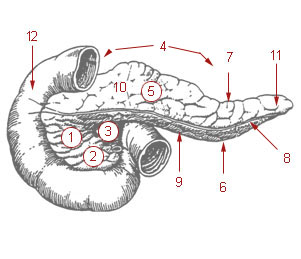
WThe pancreatic notch is a separation between the neck of pancreas and the uncinate process of pancreas.
 W
WIn medicine, a Phrygian cap is the folded portion of some gallbladders that resembles the Phrygian cap. It is a normal anatomical variant seen in 1-6% of patients. It is caused by a fold in the gallbladder where the gallbladder fundus joins the gallbladder body. Apart from the chance of being mistaken for stones on a sonogram, it has no other medical implications nor does it predispose one to other diseases. However, due to potential decrease in bile flow, it may warrant a preventive removal of the gallbladder.
 W
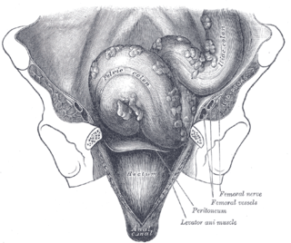
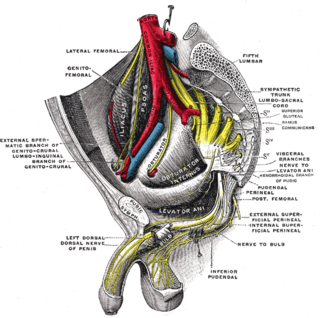
WIn human anatomy, the presacral space is inside the pelvis, behind the rectum and in front of the coccyx and sacrum. Normally it is empty, or it contains a pocket of fat.
 W
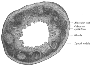
WThe Solitary lymphatic nodules are structures found in the small intestine and large intestine.
 W
WThe sphincter of Oddi, abbreviated as SO, is a muscular valve that in some animals, including humans, controls the flow of digestive juices through the ampulla of Vater into the second part of the duodenum. It is named after Ruggero Oddi. The sphincter of Oddi is relaxed by the hormone cholecystokinin via vasoactive intestinal peptide.
 W
WThe sublingual papilla or sublingual fold is a small fold of soft tissue located on each side of the frenulum linguae binding the lips to the gums within the mouth.
 W
WThe tela subserosa is a thin layer of tissue in the walls of various organs. It is a layer of connective tissue between the muscular layer and the serosa.