 W
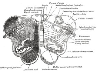
WThe medulla oblongata or simply medulla is a long stem-like structure which makes up the lower part of the brainstem. It is anterior and partially inferior to the cerebellum. It is a cone-shaped neuronal mass responsible for autonomic (involuntary) functions, ranging from vomiting to sneezing. The medulla contains the cardiac, respiratory, vomiting and vasomotor centers, and therefore deals with the autonomic functions of breathing, heart rate and blood pressure as well as the sleep wake cycle.
 W
WThe accessory cuneate nucleus is located lateral to the cuneate nucleus in the medulla oblongata at the level of the sensory decussation.
 W
WThe anterior external arcuate fibers vary as to their prominence: in some cases they form an almost continuous layer covering the medullary pyramids and olivary body, while in other cases they are barely visible on the surface.
 W
WThe anterior median fissure contains a fold of pia mater, and extends along the entire length of the medulla oblongata: It ends at the lower border of the pons in a small triangular expansion, termed the foramen cecum.
 W
WThe anterolateral sulcus is a sulcus on the side of the medulla oblongata between the olive and pyramid. The rootlets of the hypoglossal nerve emerge from this sulcus.
 W
WIn the medulla oblongata, the arcuate nucleus is a group of neurons located on the anterior surface of the medullary pyramids. These nuclei are the extension of the pontine nuclei. They receive fibers from the corticospinal tract and send their axons through the anterior external arcuate fibers and medullary striae to the cerebellum via the inferior cerebellar peduncle.
 W
WThe area postrema, a paired structure in the medulla oblongata of the brainstem, is a circumventricular organ having permeable capillaries and sensory neurons that enable its dual role to detect circulating chemical messengers in the blood and transduce them into neural signals and networks. Its position adjacent to the bilateral nuclei of the solitary tract and role as a sensory transducer allow it to integrate blood-to-brain autonomic functions. Such roles of the area postrema include its detection of circulating hormones involved in vomiting, thirst, hunger, and blood pressure control.
 W
WClimbing fibers are the name given to a series of neuronal projections from the inferior olivary nucleus located in the medulla oblongata.
 W
WThe cochlear nuclear (CN) complex comprises two cranial nerve nuclei in the human brainstem, the ventral cochlear nucleus (VCN) and the dorsal cochlear nucleus (DCN). The ventral cochlear nucleus is unlayered whereas the dorsal cochlear nucleus is layered. Auditory nerve fibers, fibers that travel through the auditory nerve carry information from the inner ear, the cochlea, on the same side of the head, to the nerve root in the ventral cochlear nucleus. At the nerve root the fibers branch to innervate the ventral cochlear nucleus and the deep layer of the dorsal cochlear nucleus. All acoustic information thus enters the brain through the cochlear nuclei, where the processing of acoustic information begins. The outputs from the cochlear nuclei are received in higher regions of the auditory brainstem.
 W
WThe dorsal nucleus of vagus nerve is a cranial nerve nucleus for the vagus nerve in the medulla that lies ventral to the floor of the fourth ventricle. It mostly serves parasympathetic vagal functions in the gastrointestinal tract, lungs, and other thoracic and abdominal vagal innervations. The cell bodies for the preganglionic parasympathetic vagal neurons that innervate the heart reside in the nucleus ambiguus.
 W
WThe inferior olivary nucleus (ION), is a structure found in the medulla oblongata underneath the superior olivary nucleus. In vertebrates, the ION is known to coordinate signals from the spinal cord to the cerebellum to regulate motor coordination and learning. These connections have been shown to be tightly associated, as degeneration of either the cerebellum or the ION results in degeneration of the other.
 W
WThe nucleus ambiguus is a group of large motor neurons, situated deep in the medullary reticular formation named by Jacob Clarke. The nucleus ambiguus contains the cell bodies of neurons that innervate the muscles of the soft palate, pharynx, and larynx which are associated with speech and swallowing. As well as motor neurons, the nucleus ambiguus contains preganglionic parasympathetic neurons which innervate postganglionic parasympathetic neurons in the heart.
 W
WThe nucleus raphe magnus, is located directly rostral to the nucleus raphe obscurus, and receives input from the spinal cord and cerebellum.
 W
WThe nucleus raphe obscurus, despite the implications of its name, has some very specific functions and connections of afferent and efferent nature. The nucleus raphes obscurus projects to the cerebellar lobes VI and VII and to crus II along with the nucleus raphe pontis.
 W
WThe nucleus raphe pallidus receives afferent connections from the periaqueductal gray, the Paraventricular nucleus of hypothalamus, central nucleus of the amygdala, lateral hypothalamic area, and parvocellular reticular nucleus.
 W
WThe obex is the point in the human brain at which the fourth ventricle narrows to become the central canal of the spinal cord.
 W
WIn anatomy, the olivary bodies or simply olives are a pair of prominent oval structures in the medulla oblongata, the lower portion of the brainstem. They contain the olivary nuclei.
 W
WThe posterior median sulcus of medulla oblongata is a narrow groove; and exists only in the closed part of the medulla oblongata; it becomes gradually shallower from below upward, and finally ends about the middle of the medulla oblongata, where the central canal expands into the cavity of the fourth ventricle.
 W
WThe accessory, vagus, and glossopharyngeal nerves correspond with the posterior nerve roots, and are attached to the bottom of a sulcus named the posterolateral sulcus.
 W
WThe respiratory center is located in the medulla oblongata and pons, in the brainstem. The respiratory center is made up of three major respiratory groups of neurons, two in the medulla and one in the pons. In the medulla they are the dorsal respiratory group, and the ventral respiratory group. In the pons, the pontine respiratory group includes two areas known as the pneumotaxic centre and the apneustic centre.
 W
WThe rostral ventromedial medulla (RVM), or ventromedial nucleus of the spinal cord, is a group of neurons located close to the midline on the floor of the medulla oblongata (myelencephalon). The rostral ventromedial medulla sends descending inhibitory and excitatory fibers to the dorsal horn spinal cord neurons. There are 3 categories of neurons in the RVM: on-cells, off-cells, and neutral cells. They are characterized by their response to nociceptive input. Off-cells show a transitory decrease in firing rate right before a nociceptive reflex, and are theorized to be inhibitory. Activation of off-cells, either by morphine or by any other means, results in antinociception. On-cells show a burst of activity immediately preceding nociceptive input, and are theorized to be contributing to the excitatory drive. Neutral cells show no response to nociceptive input.
 W
WIn the human brainstem, the solitary nucleus (SN) is a series of purely sensory nuclei forming a vertical column of grey matter embedded in the medulla oblongata. Through the center of the SN runs the solitary tract, a white bundle of nerve fibers, including fibers from the facial, glossopharyngeal and vagus nerves, that innervate the SN. The SN projects to, among other regions, the reticular formation, parasympathetic preganglionic neurons, hypothalamus and thalamus, forming circuits that contribute to autonomic regulation. Cells along the length of the SN are arranged roughly in accordance with function; for instance, cells involved in taste are located In the rostrum part, while those receiving information from cardio-respiratory and gastrointestinal processes are found in the caudal part.
 W
WThe solitary tract is a compact fiber bundle that extends longitudinally through the posterolateral region of the medulla. The solitary tract is surrounded by the nucleus of the solitary tract, and descends to the upper cervical segments of the spinal cord. It was first named by Theodor Meynert in 1872.
 W
WThe spinal accessory nucleus lies within the cervical spinal cord (C1-C5) in the posterolateral aspect of the anterior horn. The nucleus ambiguus is classically said to provide the "cranial component" of the accessory nerve.
 W
WThe spinal trigeminal nucleus is a nucleus in the medulla that receives information about deep/crude touch, pain, and temperature from the ipsilateral face. In addition to the trigeminal nerve, the facial, glossopharyngeal, and vagus nerves also convey pain information from their areas to the spinal trigeminal nucleus. Thus the spinal trigeminal nucleus receives input from cranial nerves V, VII, IX, and X.