 W
WThe temporal lobe is one of the four major lobes of the cerebral cortex in the brain of mammals. The temporal lobe is located beneath the lateral fissure on both cerebral hemispheres of the mammalian brain.
 W
WBrodmann area 20, or BA20, is part of the temporal cortex in the human brain. The region encompasses most of the ventral temporal cortex, a region believed to play a part in high-level visual processing and recognition memory.
 W
WBrodmann area 21, or BA21, is part of the temporal cortex in the human brain. The region encompasses most of the lateral temporal cortex, a region believed to play a part in auditory processing and language. Language function is left lateralized in most individuals. BA21 is superior to BA20 and inferior to BA40 and BA41.
 W
WBrodmann area 22 is a Brodmann's area that is cytoarchitecturally located in the posterior superior temporal gyrus of the brain. In the left cerebral hemisphere, it is one portion of Wernicke's area. The left hemisphere helps with generation and understanding of individual words. On the right side of the brain, it helps to discriminate pitch and sound intensity, both of which are necessary to perceive melody and prosody. Wernicke's area is active in processing language and consists of the left Brodmann area 22 and Brodmann area 40, the supramarginal gyrus.
 W
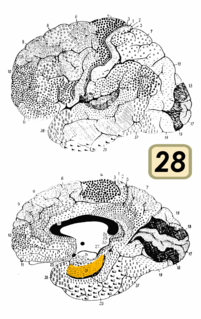
WBrodmann area 28 is a subdivision of the cerebral cortex defined on the basis of cytoarchitecture. It is located on the medial aspect of the temporal lobe and is part of the entorhinal cortex (Brodmann-1909).
 W
WBrodmann area 34 is a part of the brain.
 W
WBrodmann area 35, together with Brodmann area 36, comprise the perirhinal cortex. They are cytoarchitecturally defined temporal regions of the cerebral cortex.
 W
WBrodmann area 37, or BA37, is part of the temporal cortex in the human brain. It contains the fusiform gyrus which in turn contains the fusiform face area, an area important for the recognition of faces.
 W
WBrodmann area 38, also BA38 or temporopolar area 38 (H), is part of the temporal cortex in the human brain. BA 38 is at the anterior end of the temporal lobe, known as the temporal pole.
 W
WBrodmann areas 41 and 42 are parts of the primary auditory cortex.
 W
WBrodmann area 52 (H) or parainsular area, is a subdivision of the cytoarchitecturally defined temporal region of the cerebral cortex in the brain.
 W
WFrontotemporal dementia (FTD), or frontotemporal neurocognitive disorder encompasses several types of dementia involving the frontal and temporal lobes. FTDs are broadly presented as behavioral or language disorders. The three main subtypes or variant syndromes are a behavioral variant (bvFTD) previously known as Pick's disease, and two variants of primary progressive aphasia – semantic variant (svPPA), and nonfluent variant (nfvPPA). Two rare distinct subtypes of FTD are neuronal intermediate filament inclusion disease (NIFID), and basophilic inclusion body disease. Other related disorders include corticobasal syndrome and FTD with amyotrophic lateral sclerosis (ALS) FTD-ALS also called FTD-MND.
 W
WFrontotemporal lobar degeneration (FTLD) is a pathological process that occurs in frontotemporal dementia. It is characterized by atrophy in the frontal lobe and temporal lobe of the brain, with sparing of the parietal and occipital lobes.
 W
WThe fusiform face area (FFA); is a part of the human visual system that is specialized for facial recognition. It is located in the inferior temporal cortex (IT), in the fusiform gyrus.
 W
WThe fusiform gyrus, also known as the lateral occipitotemporal gyrus, is part of the temporal lobe and occipital lobe in Brodmann area 37. The fusiform gyrus is located between the lingual gyrus and parahippocampal gyrus above, and the inferior temporal gyrus below. Though the functionality of the fusiform gyrus is not fully understood, it has been linked with various neural pathways related to recognition. Additionally, it has been linked to various neurological phenomena such as synesthesia, dyslexia, and prosopagnosia.
 W
WThe inferior temporal gyrus is one of three gyri of the temporal lobe and is located below the middle temporal gyrus, connected behind with the inferior occipital gyrus; it also extends around the infero-lateral border on to the inferior surface of the temporal lobe, where it is limited by the inferior sulcus. This region is one of the higher levels of the ventral stream of visual processing, associated with the representation of objects, places, faces, and colors. It may also be involved in face perception, and in the recognition of numbers.
 W
WThe inferior surface of the temporal lobe is concave, and is continuous posteriorly with the tentorial surface of the occipital lobe. It is traversed by the inferior temporal sulcus, which extends from near the occipital pole behind, to within a short distance of the temporal pole in front, but is frequently subdivided by bridging gyri.
 W
WMiddle temporal gyrus is a gyrus in the brain on the Temporal lobe. It is located between the superior temporal gyrus and inferior temporal gyrus.
 W
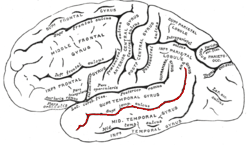
WThe superior temporal gyrus (STG) is one of three gyri in the temporal lobe of the human brain, which is located laterally to the head, situated somewhat above the external ear.
 W
WThe superior temporal sulcus (STS) is the sulcus separating the superior temporal gyrus from the middle temporal gyrus in the temporal lobe of the brain. A sulcus is a deep groove that curves into the largest part of the brain, the cerebrum, and a gyrus is the a ridge that curves outward of the cerebrum.
WTemporal lobe epilepsy (TLE) is a chronic disorder of the nervous system characterized by recurrent, unprovoked focal seizures that originate in the temporal lobe of the brain and last about one or two minutes. TLE is the most common form of epilepsy with focal seizures. A focal seizure in the temporal lobe may spread to other areas in the brain when it may become a focal to bilateral seizure.
 W
WIn the human nervous system the temporopontine fibers, a component of the corticopontine tract are lateral to the cerebrospinal fibers; they originate in the temporal lobe and end in the nuclei pontis.
 W
WThe transverse temporal gyri, also called Heschl's gyri or Heschl's convolutions, are gyri found in the area of primary auditory cortex buried within the lateral sulcus of the human brain, occupying Brodmann areas 41 and 42. Transverse temporal gyri are superior to and separated from the planum temporale by Heschl’s sulcus. Transverse temporal gyri are found in varying numbers in both the right and left hemispheres of the brain and one study found that this number is not related to the hemisphere or dominance of hemisphere studied in subjects. Transverse temporal gyri can be viewed in the sagittal plane as either an omega shape or a heart shape.
 W
WWernicke's area, also called Wernicke's speech area, is one of the two parts of the cerebral cortex that are linked to speech, the other being Broca's area. It is involved in the comprehension of written and spoken language, in contrast to Broca's area, which is involved in the production of language. It is traditionally thought to reside in Brodmann area 22, which is located in the superior temporal gyrus in the dominant cerebral hemisphere, which is the left hemisphere in about 95% of right-handed individuals and 60% of left-handed individuals.