 W
WThe basal ganglia are a group of subcortical nuclei, of varied origin, in the brains of vertebrates. In humans, and other primates there are some differences, mainly in the division of the globus pallidus into an external and internal region, and in the division of the striatum. The basal ganglia are situated at the base of the forebrain and top of the midbrain. Basal ganglia are strongly interconnected with the cerebral cortex, thalamus, and brainstem, as well as several other brain areas. The basal ganglia are associated with a variety of functions, including control of voluntary motor movements, procedural learning, habit learning, eye movements, cognition, and emotion.
 W
WThe caudate nucleus is one of the structures that make up the corpus striatum, which is a component of the basal ganglia. While the caudate nucleus has long been associated with motor processes due to its role in Parkinson's disease, it plays important roles in various other nonmotor functions as well, including procedural learning, associative learning and inhibitory control of action, among other functions. The caudate is also one of the brain structures which compose the reward system and functions as part of the cortico–basal ganglia–thalamic loop.
 W
WThe direct pathway, sometimes known as the direct pathway of movement, is a neural pathway within the central nervous system (CNS) through the basal ganglia which facilitates the initiation and execution of voluntary movement. It works in conjunction with the indirect pathway. Both of these pathways are part of the cortico-basal ganglia-thalamo-cortical loop.
 W
WThe external globus pallidus combines with the internal globus pallidus (GPi) to form the globus pallidus, an anatomical subset of the basal ganglia. Globus pallidus means "pale globe" in Latin, indicating its appearance. The external globus pallidus is the segment of the globus pallidus that is relatively further (lateral) from the midline of the brain.
 W
WThe globus pallidus (GP), also known as paleostriatum or dorsal pallidum, is a subcortical structure of the brain. It consists of two adjacent segments, one external, known in rodents simply as the globus pallidus, and one internal, known in rodents as the entopeduncular nucleus. It is part of the telencephalon, but retains close functional ties with the subthalamus in the diencephalon – both of which are part of the extrapyramidal motor system. The globus pallidus is a major component of the basal ganglia, with principal inputs from the striatum, and principal direct outputs to the thalamus and the substantia nigra. The latter is made up of similar neuronal elements, has similar afferents from the striatum, similar projections to the thalamus, and has a similar synaptology. Neither receives direct cortical afferents, and both receive substantial additional inputs from the intralaminar thalamus.
WThe indirect pathway, sometimes known as the indirect pathway of movement, is a neuronal circuit through the basal ganglia and several associated nuclei within the central nervous system (CNS) which helps to prevent unwanted muscle contractions from competing with voluntary movements. It operates in conjunction with the direct pathway.
WThe internal globus pallidus and the external globus pallidus (GPe) make up the globus pallidus. The GPi is one of the output nuclei of the basal ganglia. The GABAergic neurons send their axons to the ventral anterior nucleus (VA) and the ventral lateral nucleus (VL) in the dorsal thalamus, to the centromedian complex, and to the pedunculopontine complex.
 W
WThe islands of Calleja are a group of neural granule cells located within the ventral striatum in the brains of most animals. This region of the brain is part of the limbic system, where it aids in the reinforcing effects of reward-like activities. Within most species, the islands are specifically located within the olfactory tubercle; however, in primates, these islands are located within the nucleus accumbens, the reward center of the brain, since the olfactory tubercle has practically disappeared in the brains of primates. Both of these structures have been implicated in the processing of incentives as well as addictions to drugs. Projections to and from the islands supplement this knowledge with their involvement in the reward pathways for both cocaine and amphetamines.
 W
WThe lentiform nucleus, or lenticular nucleus, comprises the putamen and the globus pallidus within the basal ganglia. With the caudate nucleus, it forms the striatum. It is a large, lens-shaped mass of gray matter just lateral to the internal capsule. Increased volume of the lentiform nuclei has been observed in obsessive-compulsive disorder, with decreased volume conversely observed in other anxiety disorders.
 W
WMedium spiny neurons (MSNs), also known as spiny projection neurons (SPNs), are a special type of GABAergic inhibitory cell representing 95% of neurons within the human striatum, a basal ganglia structure. Medium spiny neurons have two primary phenotypes : D1-type MSNs of the direct pathway and D2-type MSNs of the indirect pathway. Most striatal MSNs contain only D1-type or D2-type dopamine receptors, but a subpopulation of MSNs exhibit both phenotypes.
 W
WThe nucleus accumbens is a region in the basal forebrain rostral to the preoptic area of the hypothalamus. The nucleus accumbens and the olfactory tubercle collectively form the ventral striatum. The ventral striatum and dorsal striatum collectively form the striatum, which is the main component of the basal ganglia. The dopaminergic neurons of the mesolimbic pathway project onto the GABAergic medium spiny neurons of the nucleus accumbens and olfactory tubercle. Each cerebral hemisphere has its own nucleus accumbens, which can be divided into two structures: the nucleus accumbens core and the nucleus accumbens shell. These substructures have different morphology and functions.
 W
WThe putamen is a round structure located at the base of the forebrain (telencephalon). The putamen and caudate nucleus together form the dorsal striatum. It is also one of the structures that compose the basal nuclei. Through various pathways, the putamen is connected to the substantia nigra, the globus pallidus, the claustrum, and the thalamus, in addition to many regions of the cerebral cortex. A primary function of the putamen is to regulate movements at various stages and influence various types of learning. It employs GABA, acetylcholine, and enkephalin to perform its functions. The putamen also plays a role in degenerative neurological disorders, such as Parkinson's disease.
 W
WThe striatopallidal fibres, also Wilson's pencils, pencil fibres of Wilson, and pencils of Wilson, are prominent myelinated fibres that connect the striatum to the globus pallidus.
 W
WThe striatum, or corpus striatum, is a nucleus in the subcortical basal ganglia of the forebrain. The striatum is a critical component of the motor and reward systems; receives glutamatergic and dopaminergic inputs from different sources; and serves as the primary input to the rest of the basal ganglia.
 W
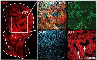
WThe striosomes are one of two complementary chemical compartments within the striatum that can be visualized by staining for immunocytochemical markers such as acetylcholinesterase, enkephalin, substance P, limbic system-associated membrane protein (LAMP), AMPA receptor subunit 1 (GluR1), dopamine receptor subunits, and calcium binding proteins. Striosomal abnormalities have been associated with neurological disorders, such as mood dysfunction in Huntington's disease, though their precise function remains unknown. Striosomes were discovered by Ann Graybiel in 1978 using acetylcholinesterase histochemistry.
 W
WThe substantia nigra (SN) is a basal ganglia structure located in the midbrain that plays an important role in reward and movement. Substantia nigra is Latin for "black substance", reflecting the fact that parts of the substantia nigra appear darker than neighboring areas due to high levels of neuromelanin in dopaminergic neurons. Parkinson's disease is characterized by the loss of dopaminergic neurons in the substantia nigra pars compacta.
 W
WThe subthalamic nucleus is a small lens-shaped nucleus in the brain where it is, from a functional point of view, part of the basal ganglia system. In terms of anatomy, it is the major part of the subthalamus. As suggested by its name, the subthalamic nucleus is located ventral to the thalamus. It is also dorsal to the substantia nigra and medial to the internal capsule. It was first described by Jules Bernard Luys in 1865, and the term corpus Luysi or Luys' body is still sometimes used.
WThe subthalamus or prethalamus is a part of the diencephalon. Its most prominent structure is the subthalamic nucleus. The subthalamus connects to the globus pallidus, a basal nucleus of the telencephalon.