 W
WThe arcuate uterus is a form of a uterine anomaly or variation where the uterine cavity displays a concave contour towards the fundus. Normally the uterine cavity is straight or convex towards the fundus on anterior-posterior imaging, but in the arcuate uterus the myometrium of the fundus dips into the cavity and may form a small septation. The distinction between an arcuate uterus and a septate uterus is not standardized.
 W
WA bicornuate uterus or bicornate uterus, is a type of mullerian anomaly in the human uterus, where there is a deep indentation at the fundus (top) of the uterus.
 W
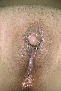
WClitoromegaly is an abnormal enlargement of the clitoris that is mostly congenital or acquired, though deliberately induced clitoris enlargement as a form of female genital body modification is achieved through various uses of anabolic steroids, including testosterone, and may also be referred to as clitoromegaly. Clitoromegaly is not the same as normal enlargement of the clitoris seen during sexual arousal.
 W
WMURCS association is a very rare developmental disorder that primarily affects the reproductive and urinary systems involving MUllerian agenesis, Renal agenesis, Cervicothoracic Somite abnormalities. It affects only females.
 W
WA rectovaginal fistula is a medical condition where there is a fistula or abnormal connection between the rectum and the vagina.
 W
WA t-shaped uterus is a type of uterine malformation wherein the uterus is shaped resembling the letter T. This is typically observed in DES-exposed women. It is recognised in the ESHRE/ESGE classification, and is associated with failed implantation, increased risk of ectopic pregnancy, miscarriage and preterm delivery. There is a surgical procedure to correct the malformation.
 W
WA unicornuate uterus represents a uterine malformation where the uterus is formed from one only of the paired Müllerian ducts while the other Müllerian duct does not develop or only in a rudimentary fashion. The sometimes called hemi-uterus has a single horn linked to the ipsilateral fallopian tube that faces its ovary.
WA uterine malformation is a type of female genital malformation resulting from an abnormal development of the Müllerian duct(s) during embryogenesis. Symptoms range from amenorrhea, infertility, recurrent pregnancy loss, and pain, to normal functioning depending on the nature of the defect.
 W
WUterus didelphys represents a uterine malformation where the uterus is present as a paired organ when the embryogenetic fusion of the Müllerian ducts fails to occur. As a result, there is a double uterus with two separate cervices, and possibly a double vagina as well. Each uterus has a single horn linked to the ipsilateral fallopian tube that faces its ovary.
 W
WVaginal anomalies are abnormal structures that are formed during the prenatal development of the female reproductive system and are rare congenital defects that result in an abnormal or absent vagina. When present, they are often found with uterine, skeletal and urinary abnormalities. This is because these structures, like the vagina, are most susceptible to disruption during crucial times of organ-genesis. Many of these defects are classified under the broader term Müllerian duct anomalies. Müllerian duct anomalies are caused by a disturbance during the embryonic time of genitourinary development. The other isolated incidents of vaginal anomalies can occur with no apparent cause. Oftentimes vaginal anomalies are part of a cluster of defects or syndromes. In addition, inheritance can play a part as can prenatal exposure to some teratogens. Many vaginal anomalies are not detected at birth because the external genitalia appear to be normal. Other organs of the reproductive system may not be affected by an abnormality of the vagina. The uterus, fallopian tubes and ovaries can be functional despite the presence of a defect of the vagina and external genitalia. A vaginal anomaly may not affect fertility. Though it depends on the extent of the vaginal defect, it is possible for conception to occur. In instances where a functional ovary exists, IVF may be successful. Functioning ovaries in a woman with a vaginal defect allows the implantation of a fertilized ovum into the uterus of an unaffected gestational carrier. A successful conception and can occur. Vaginal length varies from 6.5 to 12.5 cm. Since this is slightly shorter than older descriptions, it may impact the diagnosis of women with vaginal agenesis or hypoplasia who may unnecessarily be encouraged to undergo treatment to increase the size of the vagina. Vaginal anomalies may cause difficulties in urination, conception, pregnancy, impair sex. Psychosocial effects can also exist.