 W
WA radiologic sign is an objective indication of some medical fact that is detected by a physician during radiologic examination with medical imaging.
 W
WIn radiology, the air crescent sign is a finding on chest radiograph and computed tomography that is crescenteric and radiolucent, due to a lung cavity that is filled with air and has a round radiopaque mass. Classically, it is due to an aspergilloma, a form of aspergillosis, that occurs when the fungus Aspergillus grows in a cavity in the lung. It is also referred as Monad sign.
 W
WChondrocalcinosis or cartilage calcification is calcification in hyaline cartilage and/or fibrocartilage. It can be seen on radiography.
 W
WCrazy paving refers to a pattern seen on computed tomography of the chest, involving lobular septal thickening with variable alveolar filling. The finding is seen in pulmonary alveolar proteinosis, and other diseases. Its name comes from its resemblance to irregular paving stones, called crazy pavings.
 W
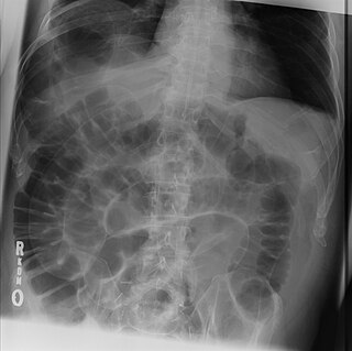
WThe cupola sign is seen on a supine chest or abdominal radiograph in the presence of pneumoperitoneum.
 W
WIn radiology, the deep sulcus sign on a supine chest radiograph is an indirect indicator of a pneumothorax. In a supine film, it appears as a deep, lucent, ipsilateral costophrenic angle within the nondependent portions of the pleural space as opposed to the apex when the patient is upright. The costophrenic angle is abnormally deepened when the pleural air collects laterally, producing the deep sulcus sign.
 W
WIn medicine, the dense artery sign or hyperdense artery sign is an increased radiodensity of an artery as seen on computer tomography (CT) scans, and is a radiologic sign of early ischemic stroke. In earlier studies of medical imaging in patients with strokes, it was the earliest sign of ischemic stroke in a significant minority of cases. Its appearance portends a poor prognosis for the patient.
 W
WIn radiology, the double bubble sign is a feature of pediatric imaging seen on radiographs or prenatal ultrasound in which two air filled bubbles are seen in the abdomen, representing two discontiguous loops of bowel in a proximal, or 'high,' small bowel obstruction. The finding is typically pathologic, and implies either duodenal atresia, duodenal web, annular pancreas, and on occasion midgut volvulus, a distinction that requires close clinical correlation and, in most cases, surgical intervention.
 W
WGround-glass opacity (GGO) is a finding seen on chest x-ray (radiograph) or computed tomography (CT) imaging of the lungs. It is typically defined as an area of hazy opacification (x-ray) or increased attenuation (CT) due to air displacement by fluid, airway collapse, fibrosis, or a neoplastic process. When a substance other than air fills an area of the lung it increases that area's density. On both x-ray and CT, this appears more grey or hazy as opposed to the normally dark-appearing lungs. Although it can sometimes be seen in normal lungs, common pathologic causes include infections, interstitial lung disease, and pulmonary edema.
 W
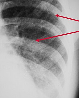
WHampton's hump, also called Hampton hump, is a radiologic sign which consists of a shallow wedge-shaped opacity in the periphery of the lung with its base against the pleural surface. It is named after Aubrey Otis Hampton, who first described it in 1940. Hampton's hump along with Westermark sign may aid in the diagnosis of pulmonary embolism, although they are rare and their sensitivities and interoperator reliabilities are low. If the sign is present in an image, there is a high chance that the person has a pulmonary embolism, but when the sign is absent a pulmonary embolism is not ruled out.
 W
WThe hilum overlay sign is an imaging appearance on chest radiographs in which the outline of the hilum can be seen at the level of a mass or collection in the mid chest. It implies that the mass is not in the middle mediastinum, and is either from anterior or posterior mediastinum(most of the masses arise from the anterior mediastinum).
 W
WThe hot nose sign refers to increased perfusion in the nasal region on nuclear medicine cerebral perfusion studies in the setting of brain death. The absent or reduced flow in the internal carotid arteries is thought to lead to increased flow within the external carotid arteries and subsequent increased perfusion in the nasal region.
 W
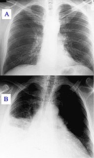
WMediastinal shift is the deviation of the mediastinal structures towards one side of the chest cavity, usually seen on chest radiograph. It indicates a severe asymmetry of intrathoracic pressures. Mediastinal shift may be caused by volume expansion on one side of the thorax, volume loss on one side of the thorax, mediastinal masses and vertebral or chest wall abnormalities. An emergent condition classically presenting with mediastinal shift is tension pneumothorax.
 W
WNotching of the ribs is a radiologic sign where the surface of the rib is deformed. It can be characterized as unilateral or bilateral, and should be differentiated between affecting the upper (superior) or lower (inferior) surface of the rib.
 W
WPeribronchial cuffing, also referred to as peribronchial thickening or bronchial wall thickening, is a radiologic sign which occurs when excess fluid or mucus buildup in the small airway passages of the lung causes localized patches of atelectasis. This causes the area around the bronchus to appear more prominent on an X-ray. It has also been described as donut sign, considering the edge is thicker, and the center contains air.
 W
WPneumatosis intestinalis is pneumatosis of an intestine, that is, gas cysts in the bowel wall. As a radiological sign it is highly suggestive for necrotizing enterocolitis. This is in contrast to gas in the intestinal lumen. In newborns, pneumatosis intestinalis is considered diagnostic for necrotizing enterocolitis, and the gas is produced by bacteria in the bowel wall. The pathogenesis of pneumatosis intestinalis is poorly understood and is likely multifactorial. PI itself is not a disease, but rather a clinical sign. In some cases, PI is an incidental finding, whereas in others, it portends a life-threatening intra-abdominal condition.
 W
WA pulmonary consolidation is a region of normally compressible lung tissue that has filled with liquid instead of air. The condition is marked by induration of a normally aerated lung. It is considered a radiologic sign. Consolidation occurs through accumulation of inflammatory cellular exudate in the alveoli and adjoining ducts. The liquid can be pulmonary edema, inflammatory exudate, pus, inhaled water, or blood. Consolidation must be present to diagnose pneumonia: the signs of lobar pneumonia are characteristic and clinically referred to as consolidation.
 W
WA ring-enhancing lesion is an abnormal radiologic sign on MRI or CT scans obtained using radiocontrast. On the image, there is an area of decreased density surrounded by a bright rim from concentration of the enhancing contrast dye. This enhancement may represent breakdown of the blood-brain barrier and the development of an inflammatory capsule. This can be a finding in numerous disease states. In the brain, it can occur with an early brain abscess as well as in Nocardia infections associated with lung cavitary lesions. In patients with HIV, the major differential is between CNS lymphoma and CNS toxoplasmosis, with CT imaging being the appropriate next step to differentiate between the two conditions.
WIn radiology, the silhouette sign refers to the loss of normal borders between thoracic structures. It is usually caused by an intrathoracic radiopaque mass that touches the border of the heart or aorta. In other words, it is difficult to make out the borders of a particular structure - normal or otherwise - because it is next to another dense structure, both of which will appear white on a standard X-ray. It may occur, for example, in right middle lobe syndrome, where the right heart margin is obscured, and in right lower lobe pneumonia, where the border of the diaphragm on the right side is obscured, while the right heart margin remains distinct. Silhouette sign is very useful in localizing lung lesions as all structures forming cardiac silhouette are in contact with a specific portion of the lung.
 W
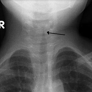
WIn radiology, the steeple sign is a radiologic sign found on a frontal neck radiograph where subglottic tracheal narrowing produces the shape of a church steeple within the trachea itself. The presence of the steeple sign supports a diagnosis of croup, usually caused by paramyxoviruses. it can also be defined as the replacement of the usual squared-shoulder appearance of the subglottic area by cone shaped narrowing just distal to the vocal cords. This is called the steeple or pencil-point sign.
 W
WIn radiology, the Terry Thomas sign is a scapholunate ligament dissociation on an anteroposterior view of the wrist. Most commonly a result of a fall on the outstretched hand (FOOSH), the scapholunate ligament ruptures resulting in separation of the lunate and scaphoid bones. This burst causes the scaphoid bone to dorsally rotate. A gap of more than 3mm is pathognomonic for scapholunate dissociation.
 W
WIn radiology, the thumbprint sign, or thumbprinting, is a radiologic sign found on a radiograph that suggests the diagnosis of either epiglottitis or intestinal ischemia.
 W
WIn radiology, the tree-in-bud sign is a finding on a CT scan that indicates some degree of airway obstruction.The tree-in-bud sign is a nonspecific imaging finding that implies impaction within bronchioles, the smallest airway passages in the lung. The differential for this finding includes malignant and inflammatory etiologies, either infectious or sterile. This includes fungal infections, mycobacterial infections such as tuberculosis or mycobacterium avium intracellulare, bronchopneumonia, chronic aspiration pneumonia, cystic fibrosis or cellular impaction from bronchovascular spread of malignancy, as can occur with breast cancer, leukemia or lymphoma. It also includes lung manifestations of autoimmune diseases such as Sjögren syndrome or rheumatoid arthritis.